


Case Report
Metastasis of Hepatocellular Carcinoma to the Maxillary Sinus
Department of Otorhinolaryngology and Head and Neck Surgery, St. Anne´s University Hospital and Faculty of Medicine, Masaryk University Brno, Czech Republic
*Corresponding author: Zuzana Horáková, Department of Head and Neck Surgery, St. Anne´s University Hospital and Faculty of Medicine, Masaryk University Brno, Czech Republic, E-mail: zuhorakova@seznam.cz
Received: April 14, 2018 Accepted: April 26, 2018 Published: May 4, 2018
Citation: Horáková Z, Binková H, Veselý K, Pažourková M, Kostřica R. Metastasis of Hepatocellular Carcinoma to the Maxillary Sinus. Madridge J Case Rep Stud. 2018; 2(1): 32-34. doi: 10.18689/mjcrs-1000108
Copyright: © 2018 The Author(s). This work is licensed under a Creative Commons Attribution 4.0 International License, which permits unrestricted use, distribution, and reproduction in any medium, provided the original work is properly cited.
Summary
Metastasis to the sinonasal region originating from a subclavicular tumour is a rare pathology, and, in the case of hepatocellular cancer (HCC), a very exceptional one. We report on a patient who presented himself with symptoms of a huge tumourous mass in the maxillary sinus. Surprisingly histopatology revealed a metastasis of an HCC. This was the first symptom of a previously undiagnosed advanced HCC. Only palliative chemotherapy could be applied. The patient died after 12 months.
Essential for the diagnosis was histopatology, including immunohistochemistry as the clinical and radiological picture might resemble the common primary cancer of the sinonasal region.
Keywords: Hepatocellular carcinoma; Sinonasal metastasis; Paranasal sinus tumors.
Introduction
A hepatocellular carcinoma (HCC) is the seventh most common malignancy worldwide with a higher incidence in men (at a ratio of 5:1), mostly in their 60s and 70s. It usually develops secondary to liver cirrhosis or hepatitis B and C, (up to 80% of cases). HCC develops distant extrahepatal metastases early: 25-50%, at the time of diagnostics. This is the poorest prognostic marker with an expected survival time less than one year.
HCC metastases occur most frequently in the lungs (55%), abdominal lymph nodes (41%) and bones (28%). The occur only very rarely in the head and neck area [1,2]. There have been published case reports of occurrence in the maxila, mandible or oral cavity, orbit, skull base or parotis [3-12]. The incidences have increased significantly during the past few years, on account of better treatment outcomes and longer survival.
Case Report
A 75-year-old male presented himself with a painful resistance under the inner corner of his right eye, which had been slowly growing for 3 months. He had a history of an asymptomatic bilateral adrenal adenoma and multiple liver lesions which was stable on CT, and thus without any further therapy, and monitoring began 5 years ago. He had lost 10 kg of weight, but otherwise was without problems. Laboratory values were long-term normal, with negative HbsAg. He suffered from practical blindness due to cataracts.
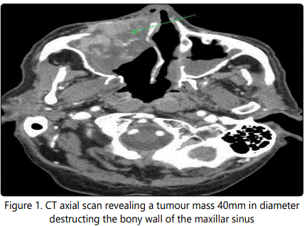
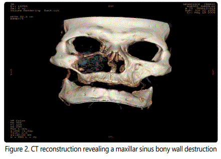
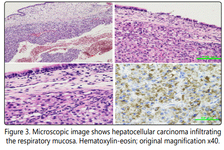
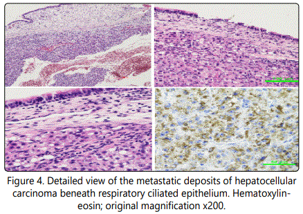
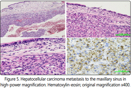
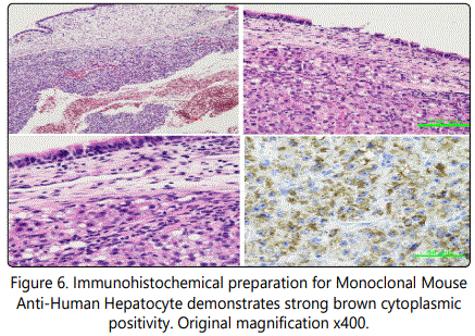
The opthamic-out patient antibiotic treatment did not result in any improvement and for progression of this resistance he was referred to ENT. A CT was done which revealed a tumour mass in the right maxillary sinus, 4x5 cm in size, destructing an anterion bony wall and invading an orbit and a nasal cavity. (Figures 1-2) Surgical exploration followed via approach secundum Denker. A macroscopically greyish-white ovoid tumoar with multiple grooves 38x29x18 mm was subtotally removed (except periorbit). Histopathologic samples were processed by the standard formol-paraffin technique and showed malignant epithelial tumour with cells growing in trabeculae, with eosinophilic finely granular cytoplasm, infiltrating the respiratory mucosa. Immunohistochemical analysis proved expression of Hepatocyte (Monoclonal Mouse Anti-Human Antibody, clone OCH1E5, Dako, USA), considered specific for hepatocellular differentiation.
Histopathological and immunohistochemical findings were consistent with metastatic hepatocellular carcinoma. (Figures 3-6)
The CT confirmed multiple liver lesions (size 50x55 mm, 3 smaller 7-18 mm), both adrenal metastases, (insize of 65x40x35mm, left 41x27x38 mm) and in mesentery (15 mm), but without evidence of lung metastases.
Only palliative chemotherapy was applied (FU weekly for 7 months), with multiple dissemination on hepatic failure. The patient died 12 months after diagnosis.
Discussion
Metastasis of HCC can disseminate to the head and neck region either via a hematogonenous or lymphatic route. Hematogenously, two ways are supposed. The first one is via the portal venous system after invading the hepatic veins, through pulmonary circle to the arteries, supraclaviculary. In these cases, pulmonary metastases always precede any further dissemination. The second way is assumed through the pre- and vertebral venous system (Batos´s plexus), due to an increased intrathoracal retrograde pressure to the jugular system, through multiple venous sinuses (plexus pterygoideus, pharyngeus, cavernosus) to the sinonasal area. In our patient, due to the absence of lung metastases, we suppose the second way. The lymphatic route passes through intrahepatic paths and the thoracic duct to the peripankreatic, celiac and paraaortal lymph nodes and retrogradely through the intercostal and mediastinal to supraclavicular vessels.
The diagnostic pitfall in sinonasal malignity is in its variable and nonspecific symptomatology, often similar to normal sinusitis (nasal obstruction, pressure and pain in the face, epistaxis). Face deformation and bulbar proptosis may appear later. Faster and more severe symptoms develop when a tumour invades the orbital structures (eyelid ptosis, eyeball proptosis, diplopia, bluered vision).
A CT is usually the first choice of radio diagnostic method which displays generally enhancing tumourous mass, possibly with osteolysis. The CT scan is mostly typical for malignancy, distinguishable from benign or inflammatory pathology but not from primary sinus carcinoma. An MRI completes information about soft tissues (metastasis is hypo intense on T1 and hyper intense on T2).
The final diagnostic decision must be based on histological results (including immunohistochemistry). Ninety-six percent of the malignancies are the primary carcinomas of the PNS or a continuously growing mass from neighbouring structures. The distant metastasis arising from the subclavicular tumor is a very rare pathology. As its radiological, as well as clinical picture is indistinguishable from the primary tumour of PNS, metastatic origin is not often considered in differential diagnosis. Even more challenging was the issue in our case when this orbital symptomatology was the first symptom of an already advanced disease with multiple extrahepatic metastases [3,5]. In a morphological histological differential diagnosis, metastasis from hepatoid carcinoma must be distinguished from another primary tumour site. The final correct diagnosis requires a complete clinico-pathological correlation.
Sinonasal metastases originate mostly from renal tumors (over 70%), less frequently from the gastrointestinal, urogenital, or lower respiratory tract, or from breast tumours. HCC metastases are rarely described; about 20 cases have been published so far [8].
Palliative chemotherapy is indicated for treatment of advanced HCC tumour with extrahepatic metastases; it can be replaced by biological or immunotherapy or only by a best supportive care in patients with poor performance status. Surgery, metastasecomy, is regarded as a palliative solution to relieve serious local symptoms. However, treatment results are not very encouraging, with short life expectancy (reported five-year survival rate 0.8% for men and 4.4% for women), between 1-21 months in the published cases. The treatment applied in our patient with a palliative intention, brought an above-average survival time of 12 months.
Conclusion
Distant metastases to sinonasal area from subclavicular tumous are very uncommon, and both clinically and radiologically are hardly distinguishable from a primary tumour. Histopatology including immunohistochemistry is essential for diagnosis. A majority of the metastases originate from renal carcinoma; a case of a hepatocellular cancer is very rare. Advanced disseminated disease is primarily diagnosed and can be treated with palliative or best supportive care only, with a short expected survival. The presented case is interesting for the atypical course of the disease, when the infiltration of the maxillary sinus developed as the first symptom of an advanced disease with multiple extra hepatic metastases.
Figures
Conflicts of interest: There is no conflict of interest
References
Contact us for any additional information - contact@madridge.org
 Madridge Publishers is licensed under a Creative Commons Attribution 4.0 International License.